

What do neurosurgeons need for successful trepanation?

The evoDrill single-use cranial perforator from evonos is the result of intensive discussions with surgeons: the skull drill allows precise trepanation with less effort and ensures fast openings.
The right perforator for modern trepanation
Skull drills used in modern trepanation need to recognize the fact that every user, patient, and clinical situation is different. We have had intensive discussions with surgeons and set ourselves the goal of solving this problem and achieving optimal results for both doctors and patients during trepanation. It has succeeded, and the result has set new standards in cranial surgery: In practice, the evoDrill has developed into one of our most sought-after products within the last ten years and is highly recognized all over the world. Precision – made in Germany, 100 percent German quality.
Trepanation is about performance, not power
This procedure became known as "Trepanation" from the Greek word "trypanon" for drill and describes in medicine an operative procedure in which a human body cavity is opened mechanically, usually by drilling with the help of a perforator. A significant proportion of efficient and successful trepanations are accounted for by the instruments used. Our powerful evoDrill perforator supports the doctor in the operation to perform a skull trephination as precisely, simply, and safely as possible. The unique design of the evoDrill is an important innovation since it requires less effort and at the same time offers more precision.
For which indications does trepanation surgery become necessary?
Trepanation surgery or "cranial perforation" refers to cranial trepanation, i.e. the surgical opening of the skullcap in living humans. There may be various reasons why a surgeon removes part of the cranium: to perform surgical interventions inside the skull, to remove projectiles that have penetrated into someone's head, or to reduce the internal cranial pressure caused by increased brain blood volume after serious cranial injury or skull fracture. In this case, one also speaks of a relief trepanation or decompression trepanation.
Which instruments contribute to the success of a skull trepanation?
A successful skull trepanation can be carried out quickly, is uncomplicated, and should not require increased effort. Our drill can meet all these requirements. Thanks to the optimized cutting geometry of the evoDrill, skulls can be opened within a very short time without effort and the bone chip can be dissipated cleanly. The opening of skulls can take the form either in the form of a hole – called perforation or trepanation – or by the temporary removal of a part of the skull bone (decompressive craniectomy).
An important advantage, which of course also comes with our perforator: the integrated auto-stop mechanism protects the Dura from injury. The benefits of this feature include ease of use and safety on the one hand and the shortest possible duration of surgery.
Skull trepanation – a standard neurosurgical procedure
A cranial trepanation or cranial perforation is considered in neurosurgery as an uncomplicated standard intervention. Catheters and drainages can be inserted via the cranial hole, for example, to relieve a space-demanding hematoma or in case of increased intracranial pressure to drain the cerebrospinal fluid.
Likewise, the trepanation holes obtained by cranial perforation facilitate access for minimally invasive surgical procedures such as neuroendoscopy. Especially for thinner bones, for example for pediatric applications, evonos offers not only the 3 mm standard product but also 1 mm variants – so our portfolio of perforators is completely tailored to individual needs.
What types of trepanation hole are there?
The main methods for the opening of the skull are either a single hole (such as that made by a circular trephine) or a sawn-out piece of bone, defined by rectangular intersecting cuts. The latter is often temporarily stored in the abdomen until reinserted, which causes it to grow back faster. In cranial trepanation, two different surgical procedures are used: In osteoplastic trepanation, the piece of skull is used again to seal the surgical wound. In the more modern osteoclastic trepanation, the closure of the resulting defect occurs in other ways for example, by implanting pieces made of titanium.
What happens after the actual trepanation or burr hole surgery?
The resulting trepanation hole is closed at the end of a neurosurgical operation in most cases. For this purpose, different methods are used: As a rule, a drill hole plate made of titanium is attached to the skull with corresponding titanium screws and covers the trepanation hole. At the same time, the plate serves to protect the underlying meninges. Another method is to fill the hole created by burr hole surgery with specialist cement, which is mixed during the operation.
Drilling holes in heads: Brain drilling made easy
Drilling holes in heads made easy. Our evoDrill perforator has many years of experience, combined with product-specific know-how. The drill is made for your requirements in skull trepanation and is 100 percent manufactured in Germany. At evonos, we believe that a perforator should be designed for brain drilling in such a way that it allows precise cuts to be simple, safe, and without much pressure. Because what always counts in the end is the result and the associated quality of life of the patient.
What are the early origins of trepanation?
Trepanation and interventions on the heads of living people are by no means a technique of modern medicine, but rather a look back reveals a millennia-old history of skull openings – even self-trepanation – among numerous peoples in South America, Africa, and Europe. Skull trepanation was already known 5,000 years ago and the evidence is detectable in many regions. By the way, research also includes the thesis that drilling a hole in the skull is the earliest surgical method and treatment that humans have used.
Exciting findings from research
Trepanned skull finds and historical literature suggest different reasons for trepanations; from scientific investigations of the skulls, one can draw conclusions about the technology used, tools, and survival rates. For example, if a skull with a hole shows traces of healing processes at the edges, this proves that the skull opening has survived. Depending on the region and period, researchers assume a survival rate of 50 to 90 percent.
From prehistoric times to Hippocrates to African medicine men
Evidence of ancient trepanation has been found in present-day Morocco; from about 10,000 BC they can be documented by European Mesolithics, from the Neolithic in East Asia, e.B. in ancient China, but also in archaeological evidence found in France. The oldest example in Western and Central Europe is a human skull dating between 5200 and 4900 BC, which was found in Alsace in 1996.
Human remains from the early Middle Ages in southwestern Germany prove medical trepanations. Skulls found in Hungary suggest that trepanation continued its long history from the 10th century onwards. Ephraim Squier's skull, discovered in Cuzco Peru in 1865, was evidence that skulls were being trepanned in South America around the 16th century.
The skulls showed evidence of ritual trepanation rather than natural cranial injuries, depressed fractures, or any attempt to address medical conditions. However, these disappeared visibly with the spread of Christianity at the beginning of the 12th century. A special case is the East African trepanations.
They only became known in Europe around the middle of the last 20th century, when British doctors photographed and published successful operations. They were able to locate more than 20 medicine men who were still making openings in skulls. The film recording of such a trepanation in 1958 also showed that surgery was carried out without narcotics. Today, operations on skulls without specialist supervision are officially prohibited in Kenya.
Why a fractured skull could be good for you
It is hard to understand the reasons for a skull trepanation. But one assumes that it was not about an operation in the brain. Rather, the skullcap was opened in order to relieve pressure on the brain through the outflow of fluid, for example after an injury. Small fragments could also be removed.
Perhaps the oldest surgical procedure involving trepanation holes is recorded by the ancient Greeks. Medical history documents reveal that, if the child's head could not pass the woman's bony pelvis, a hole was drilled into the head of these children. This was done for medical reasons in order to reduce it somewhat. Before the introduction of cesarean section, the hole in the skull was seen as the only way to save the life of the mother, although not the child. This rather extreme measure was used until the 18th century.
What human remains tell us about drilling holes in heads
In addition to medical trepanations, human remains indicate ritual-based skull openings. It is assumed that Stone Age trepanations were carried out for cultural reasons, for example, to allow evil spirits to escape through the created opening. Conversely, trepanation gave a positive spirit access to the affected person through the head wound.
As evidence for such operations, it is considered that in most finds a closure of the skull opening was missing and the removed piece was worn pierced as an amulet, often found amongst the human remains. To this day, there are some who believe that the act of trepanning can increase consciousness or address conditions such as chronic fatigue syndrome. There were also human remains found in southern Russia where skulls showed head injuries or boring holes in the same way – in the middle, above the occipital.
Due to anatomical peculiarities, this site is one of the most dangerous for a skull opening – which, in addition to the lack of evidence of skull fractures, head wounds, or diseases, suggests that these people were drilling holes in heads for ritual trepanation reasons.
Historical techniques and ritual trepanation tools from the neolithic period
Skull openings of the Stone Age are round or oval, others rectangular, square, or T-shaped (eastern France). The earliest tools used for trepanation are unknown, but the use of shells could be detected electron microscopically at the bone edges, suggesting a technique of scraping. In rectangular or square trepanations, saw marks are usually recognizable in the corners of trepanned skulls that archeologists found in human remains.
The Greek physician Hippocrates (c. 460–370 BC) used a perforative and crown saw for skull openings; in ancient times, arched drills were also used. From numerous trepanations in the 16th century, it is known that in addition to tools such as hammers, chisels, or knives, screwdrivers, or primitive drilling rigs were also used.
In total, various scraping, cutting, and drilling techniques on the head have been used as a common procedure: if the surgeons with a hard and sharp tool gradually scraped tissue away until they reached the brain, they had a higher level of control. Cuts, on the other hand, could lead to injuries.
Advocates of trepanation – trepanation between science, art, and obscurity
Interestingly, Amanda Feilding a fierce advocate of trepanation had the idea of making a documentary film about this neurosurgical procedure. The film known as The Heartbeat in the Brain was released in 1970. In it, Amanda Feilding is drilling a hole into her own forehead, piercing the skin by using a dentist’s drill. Many of the audience fainted during this scene. Thus, the art of trepanation was even discussed artistically.
Advocates of trepanation – of whom many have drilled through their own skull – have also claimed that freeing the brain of the barrier of the skull not only increases blood flow and thus, increases concentration but could also bring about a state of mind that makes one feel permanently high. People, such as Peter Halvorson, self-trepanned himself in 1972 with an electric drill in Amsterdam. Supposedly his trepanation led to heightened energy levels, focus, and drive. He founded the International Trepanation Advocacy Group in 1997.
Is trepanation still used today? A look at trepanation in modern times
Trepanation is central to modern craniotomy and craniectomy. "Craniotomy", derived from the Latin "cranium", refers to the neurosurgical opening of skulls by trepanation. This creates access into the cranial cavity in order to carry out further neurosurgical intervention on the brain, for example, the resection of a tumor. In this procedure, part of the skull is separated out. After surgery, the lid is reinserted, provided that it is not destroyed by tumor growth or trauma.
"Craniectomy" means the removal of parts or the entire skull roof. Such an operation is a modern method performed to create space for the increased volume with an increase in pressure in the skull. The removed lid is stored so that it can later be used for reimplantation. A craniectomy is indicated as a last resort if increased pressure cannot be sufficiently reduced with conservative, i.e. non-surgical measures, for example in the following indications:
- acute cerebral edema (e.g. after head trauma or stroke)
- Cerebral hemorrhage
- local or extensive cerebral inflammation
However, it is not indicated if an improvement in pressure or the underlying disease is unlikely, for example, in a malignant tumor.
What should be considered in the case of craniectomy?
When removing the skullcap, the underlying venous blood flow (sinus) must not be injured. This limits the spaces and dimensions of the removal so that practically usually a hemicraniectomy (one-sided removal of the skull roof) takes place over the affected hemisphere. Also, after the procedure, storage of the patient's head without pressure on the brain must be ensured.
In the next step, the resulting defect must be covered plastically. This is done by a dura extension plastic, i.e. a liquor-tight closure of the meninges with the inclusion of a graft (e.g. fascia, pericardium).
Preservation of the bone is possible by implantation in the abdominal cavity or by deep freezing. The disadvantage of storage in the body is a slow breakdown of substance by the immune system. In contrast, the lid is also immediately available after a transfer to another hospital. The transport of the frozen lid to its "owner" is extremely time-consuming for legal reasons. If the craniectomy can only be corrected after several weeks by replantation, a protective helmet is often necessary.
Cranioplasty – possibilities of skull defect coverage
After intensive therapy of acute brain diseases, the medical question of covering cranial defects or calottendefekts arises. The reasons for this are manifold. For example, pressure-relieving detections account for around 50 percent of cases in the event of a severe intracranial increase in pressure. Other causes include:
- Severe traumas with multiple fractures without reconstructive possibility from the body's own bone substance
- Tumor removal
- Aseptic dying bone tissue and removal after neurosurgical procedures
- Infections (local and generalized) after neurosurgical operations
- Rejections of other substitute materials
The first recommendation after craniotomy is always to cover defects in skulls with their own material, even if the infection rates of up to 25 percent are considerable. After successful implantation, a very good individual defect coverage can be achieved through bone integration.
Nevertheless, the number of patients who cannot use their own bone material is growing. For example, when these patients suffer from increased headaches, focal symptoms, epilepsy, "sinking scalp flap syndrome" and brain-organic psycho-syndromes. In the case of large defects, brain prolapse (swelling pulsating brain mass), and brain shift (brain displacement) can occur.
Often disorders such as exclusion, depressive moods, and depression also occur. In the case of skull defect coverage, functional, psychological, and aesthetic aspects are relevant. In such cases, it is important to find the optimal replacement material for each individual patient.
Choosing the right implant
For neurosurgery, various materials and manufacturers are offered in Germany. Smaller defects of less than 4 cm x 4 cm can be covered by bone replacement (calvarian split, hip), titanium nets, or PMMA. The fit is created intraoperatively.
For larger or more complex skull defects, this option is not recommended for cosmetic and functional reasons in order to achieve an optimal implant-bone transition and to ensure implant stability. Essential criteria for the choice of the optimal implant are the material and the manufacturing process because these play an important role in the fit and have a decisive influence on intraoperative handling and postoperative healing.
Why there are many reasons for evoShape
evoShape, the patient-specific skull implant from evonos, meets the highest anatomical and surgical requirements. Thanks to the development of the computer, the implant can be made CAD/CAM-based precisely to fit the cavity perfectly. The material used, polyetheretherketone (PEEK), a high-performance thermoplastic with a lower documented infection rate than PMMA and low thermal conductivity, is stable but not brittle and can be adapted with processing tools if necessary.
Entdecken Sie die Innovationen, die den Unterschied machen
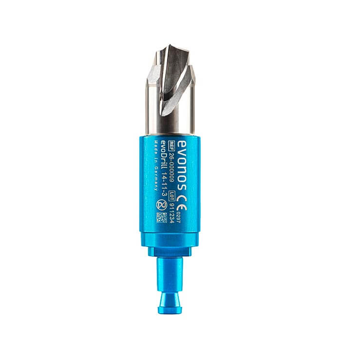 evoDrillCranial perforator
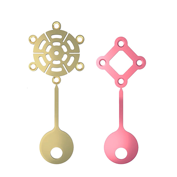 evoFixNeurosurgical osteosynthesis system
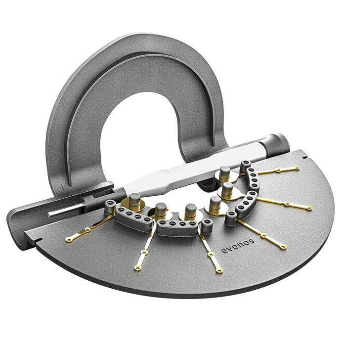 evoFix SterileOsteosynthesis system
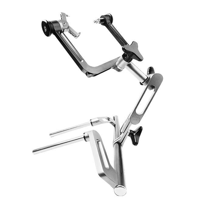 evoBaseTitanium head-positioning system
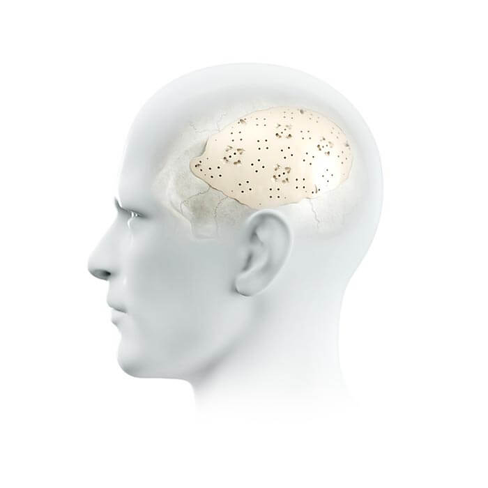 evoShapePatient-specific implants
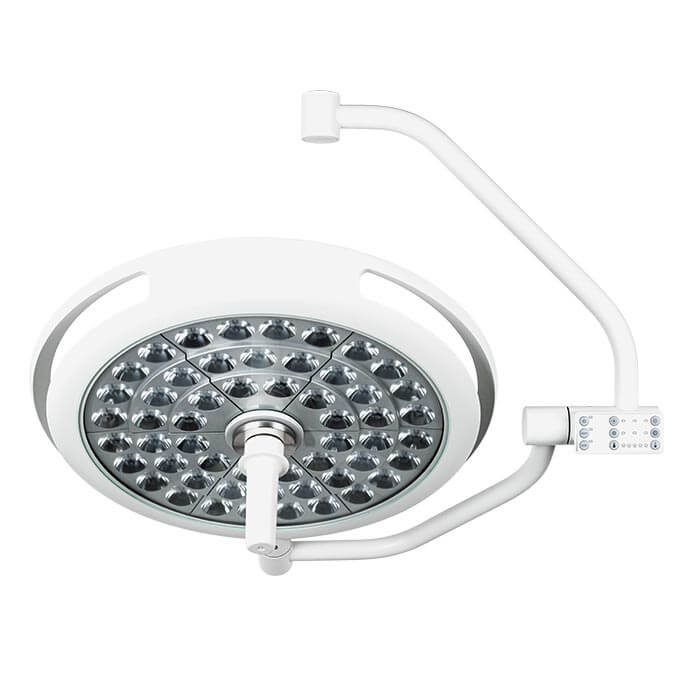 evoLightOperating room lighting system
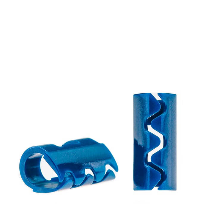 evoClipRaney scalp clips & applying forceps
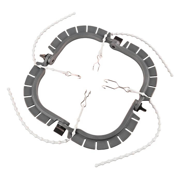 evoFrameSingle-use soft tissue retraction system
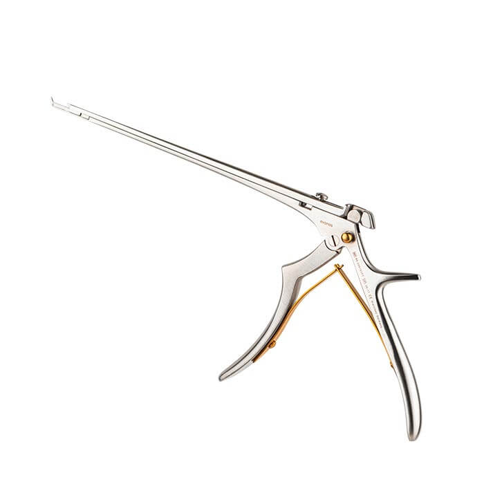 evoBiteRongeurs and laminectomy punches
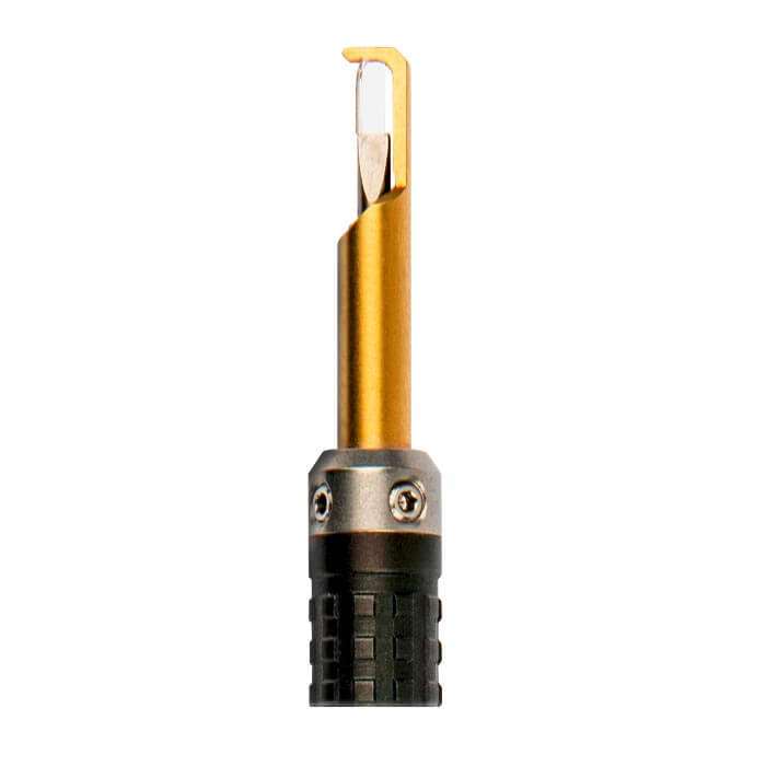 evoCaratDiamond knives for neurosurgery